MRI & MRA
Diagnostic Imaging Services

What is an MRI and how is it different from an MRA?
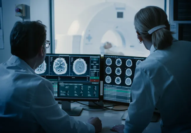
Magnetic Resonance Imaging (MRI) is a safe, painless procedure that uses a magnetic field and radio waves to produce images of body structures and organs.
Magnetic Resonance Angiography (MRA) uses MRI technology to produce images of blood vessels to detect, diagnose, and aid the treatment of heart disorders, stroke, and blood vessel diseases.

Are You Pregnant?
If you are pregnant, please discuss with your physician prior to scheduling an MRI. Please also notify the schedulers when making your appointment, as well as the technician at the imaging center.
Are You Claustrophobic?
If you are claustrophobic, please discuss with your physician prior to scheduling an MRI. If you require sedation, it must be arranged in advance with your referring physician and you will be responsible for making arrangements for someone to drive you home.
Summit's New Wide Bore MRI Scanner Featured on WATE TV's Health Watch
WATE reporter Tearsa Smith visitedSummit Diagnostic Imaging Center for a first-hand look at Summit MedicalGroup's new wide-bore MRI scanner, which provides both patients and physicianswith a variety of enhancements and benefits versus traditional or open MRIscanners.
General Test Preparation
- Pregnancy. If you think you may be pregnant, please discuss with your physician prior to scheduling and please inform the technologist.
- What to wear. Wear something comfortable with no metal, snaps, or zippers. Sweat suits are preferred. Do not wear jewelry, including body piercings.
- Medications. You may take medication as usual.
- Metal. You will not be able to enter the MRI room with any metal present on or around your body. If you have brain aneurysm clips, a pacemaker, or history of metal fragments in your eye, please inform the technologist.
If you will require sedation for this procedure, please make arrangements for someone to drive you home.
Maximum weight capacity for MRI equipment: 350 lbs.
MRI - Brain
An MRI of the brain is used to determine if you have abnormalities within your brain tissue such as cancer, tumor, stroke, or others.
Prep
Patients should refrain from using heavy eye makeup. Also, see General Prep above.
What to expect during the test
You will be placed on a narrow imaging table that will move your head into the center of the scanner. A coil will be placed over your head to send an imaging signal to the scanner and the exam will last approximately 20-30 minutes. You may be given an injection of a small amount of MRI contrast called gadolinium part way through the exam. You will be asked to remain still during the pictures. The technologist will be in contact with you during the examination. You will be given a call button that will allow you to contact the technologist during the scan.
MRI - Spine
An MRI of the spine is used to determine if you have abnormalities within your spinal column such as a herniated disc, compression fracture, spinal stenosis, cancer, tumor, infection, or others.
Prep
General Prep only - see above
What to expect during the test
You will be placed on a narrow imaging table that will move the body part being scanned into the center of the scanner. You will be lying on a coil that sends an imaging signal to the scanner and the exam will last approximately 30-40 minutes. You may be given an injection of a small amount of MRI contrast called gadolinium part way through exam. You will be asked to remain still during the pictures. The technologist will be in contact with you during the examination. You will be given a call button that will allow you to contact the technologist during the scan.
MRI - Abdominal Region
An MRI of the abdominal region is used to determine if you have abnormalities within your abdomen such as lesions, tumors, cancer or infection, or others.
Prep
Do not eat or drink anything for 4 hours prior to the test. Also, see General Prep above.
What to expect during the test
You will be placed on a narrow imaging table that will move your abdomen into the center of the scanner. A coil will be placed over your abdomen to send an imaging signal to the scanner and the exam will last approximately 30-40 minutes. The technologist will be monitoring your breathing and will be giving you breathing instructions throughout the exam. You may be given an injection of a small amount of MRI contrast called gadolinium part way through the exam. You will be asked to remain still during the pictures. The technologist will be in contact with you during the examination. You will be given a call button that will allow you to contact the technologist during the scan.
MRI - Extremity
An MRI of the extremity joint or non-joint is used to determine if you have abnormalities within your hip, knee, ankle, foot, thigh, shoulder, elbow, or wrist such as torn tendons or ligaments, bone abnormalities, torn muscle tissue, or others.
Prep
General Prep - see above
What to expect during the test
You will be placed on a narrow imaging table that will move the body part being scanned into the center of the scanner. A coil will be placed over your joint or non-joint area to send an imaging signal to the scanner and the exam will last approximately 30-40 minutes. You will be asked to remain still during the pictures. The technologist will be in contact with you during the examination. You will be given a call button that will allow you to contact the technologist during the scan.
MRA - Brain or neck
An MRA brain or neck is used to determine if you have abnormalities within your arteries such as an aneurysm, stenosis or blockage, or others.
Prep
General Prep only - see above
What to expect during the test
You will be placed on a narrow imaging table that will move your head or neck into the center of the scanner. A coil will be placed over your head or neck to send an imaging signal to the scanner and the exam will last approximately 20-30 minutes. The radiologist may give you an injection of a small amount of MRI contrast called gadolinium part way through the exam. You will be asked to remain still during the pictures. The technologist will be in contact with you during the examination. You will be given a call button that will allow you to contact the technologist during the scan.
MRA - Abdominal Region or Lower Extremity
An MRA of the abdominal region or lower extremity run-off is used to determine if you have abnormalities within your arteries such as an aneurysm, narrowing, blockage, or others.
Prep
Do not eat or drink anything for 4 hours prior to the test. Also, see General Prep above.
What to expect during the test
You will be placed on a narrow imaging table that will move your abdomen into the center of the scanner. A small I.V. will be inserted into your arm through which you will be given an injection of a small amount of MRI contrast called gadolinium part way though the exam. For an abdominal scan a coil will be placed over your abdomen to send an imaging signal to the scanner, and the exam will last approximately 20-30 minutes. For a lower extremity run-off scan you will be lying on a coil that sends an imaging signal to the scanner, and the exam will last approximately 30-40 minutes. The technologist will be monitoring your breathing and will be giving you breathing instructions throughout the exam. You will be asked to remain still during the pictures. The technologist will be in contact with you during the examination. You will be given a call button that will allow you to contact the technologist during the scan.
